
Do you think this is a genetic mutation that gives you special abilities such as “automatic magnification when looking at things” and “automatic deformation when looking at things”? no It is very likely that there is a “membrane” in the macula, and from an ophthalmic clinical perspective, it is the anterior macular membrane that has come to you.
What is macula? What is macular membrane?
The macula itself is not a disease, it is an important area responsible for central vision, located in the center of the retina. It is also the most densely populated area of the optic nerve photoreceptor cells, mainly responsible for fine vision and color recognition. Generally, the visual examination of the human eye is to check the visual ability of the macula area. Macular membrane is one of the common macular diseases.

What is macular membrane?
The anterior macular membrane is a fibrous membrane that grows along the surface of the limiting membrane in the macular area of the retina. Simply put, it is a “membrane” that grows in front of the macula.
According to clinical data, about 6.4% of people over 50 years old suffer from macular membrane, with about 20% suffering from bilateral diseases.
What are the causes of macular membrane?
The etiology of macular membrane can be divided into primary and secondary types
The primary cause is mainly due to eye degeneration, just like wrinkles appearing on old skin, which is more common in adults over the age of 50, especially in patients who have experienced posterior vitreous detachment. Therefore, macular membrane is considered one of the age-related degenerative diseases.
Secondary diseases are usually caused by intraocular inflammation or other vascular diseases, such as deep myopia, diabetes retinopathy, retinal vascular diseases, etc. In addition, retinal tear or detachment, inflammatory reaction at the back of the eye, surgical trauma, eye trauma, etc. have occurred, which may cause the macular membrane.
Symptoms of anterior macular membrane
The macular membrane can cause distortion and edema of the retina in the macular area, leading to symptoms such as decreased vision, distorted vision, and enlarged vision in patients.
In the early stage of macular membrane, it is manifested as enhanced reflection on the surface of the retina, resembling a layer of glass paper; In the later stage of the macular membrane, it appears as a semi transparent or opaque gray white membrane, accompanied by macular folds, tortuous blood vessels, macular displacement, and retinal hemorrhage.
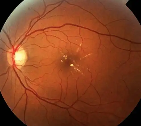
In the fundus photo of patients with mid-term macular membrane, the translucent fibrous membrane (blue arrow) is involved in the retinal blood vessels, which are straightened, twisted, and deformed.
When the macular membrane develops to the middle and late stages, severe damage to the macula in the fundus will have irreversible effects on vision.
Treatment of anterior macular membrane
The key to the treatment of macular membrane lies in early detection and timely surgery. Macular membrane surgery can be divided into three steps:
Step 1: Use a miniature vitrectomy device to remove the vitreous body. (See Figure 1)
Step 2: Place micro forceps into the eye and directly peel off the anterior membrane.
Step 3: Stain the inner membrane with a special dye, and then peel it off with micro forceps. (See Figure 2)
Due to the varying thickness and texture of the macular membrane, doctors may encounter different situations when removing it: some patients have a very thin macular membrane that can be easily removed by gently covering the macular spot; Some patients’ macular membrane is very fragile and needs to be gradually peeled off, which tests the doctor’s skills and patience; Some patients also have a tight adhesion of the macular membrane to the macular spot, and doctors need to carefully and cleverly peel it off to avoid damaging the macular spot.
Therefore, verbally saying ‘tear off the front membrane’ may sound very easy, but in actual surgery, it is quite complex and tests the skills and endurance of the surgeon.
Xima Ophthalmology is equipped with internationally leading fundus examination instruments such as optical coherence tomography (OCT), and has a strong and professional team of international fundus doctors, making macular degeneration and other fundus diseases a characteristic specialty of Xima Ophthalmology. Many macular degeneration surgeries that cannot be performed in ordinary ophthalmic hospitals can achieve very good treatment results at Xima Ophthalmology.
Special Notice
The key to treating macular membrane lies in early detection and surgery, and the early symptoms of macular membrane are similar to those of presbyopia, both of which are vision loss and blurred vision. Many middle-aged and elderly people mistakenly believe that they are suffering from presbyopia and delay treatment, thereby increasing the difficulty of surgical treatment in the later stage.