
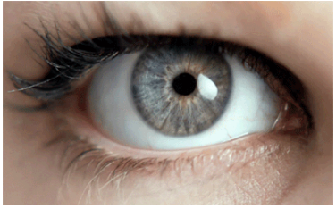
The eyes are a wonderful organ that can help people receive various visual information from the outside world, allowing them to understand the world they live in. It is precisely because of the existence of the eyes that people’s lives become colorful and vibrant.
However, as one of the precise and complex physiological organs in the human body, the eyes are also very fragile.
When people have healthy eyes, they often do not realize the importance of their eyes. Only when their vision problems occur do they realize how precious their eyes are. Therefore, we must always take care of our eyes and prevent various eye diseases. In clinical practice, there are four groups of people who are prone to eye diseases and need regular eye examinations to prevent the occurrence of eye diseases.
- Middle aged and elderly people
In middle and old age, the metabolism of the body slows down, and various functions of the body gradually decline, making it easy to develop some diseases. Among them, ophthalmic diseases are more obvious and directly reflected in vision, thereby affecting the quality of life of middle-aged and elderly people in their later years.
Ophthalmologists remind middle-aged and elderly people to be vigilant about age-related eye diseases such as cataracts, presbyopia, eye stroke, age-related macular degeneration, etc.
These eye diseases will increase the incidence rate with age. If you develop the habit of checking your eyesight regularly, you may find and treat them as soon as possible.
This not only achieves better therapeutic effects, but also has the potential to prevent and control further deterioration of diseases, allowing elderly eye diseases to be prevented before they occur.
- Patients with “three highs”
Many people believe that diabetes, hypertension and hyperlipidemia will only damage the heart, brain and blood vessels, and will not affect vision health. But in reality, these three chronic diseases will cause chronic damage to the fundus of the eyes over time.
Ophthalmologists point out that patients with “three highs” are more likely to develop eye diseases due to two factors.
One is that the “three highs” belong to systemic diseases. Poor control of the condition can affect the eyes at different stages.
For example, hypertension and hyperlipidemia can cause retinal artery or vein blockage, optic neuropathy and other eye diseases, and diabetes can cause a series of eye diseases such as diabetic retinopathy.
Secondly, drugs used to treat the “three highs” can also cause damage to the eyes.
Many cardiovascular disease treatment drugs have been clinically proven to have certain effects on the eyes, and long-term use of such drugs can cause varying degrees of damage to the eyes.
Therefore, patients with “three highs” should not take it lightly. These factors will gradually cause greater damage to the eyes over time. It is recommended that “three highs” patients undergo regular eye examinations to prevent the occurrence of “three highs” eye diseases.
- Highly myopic individuals
Due to the longer axial length of high myopia (axial myopia) compared to normal individuals, it is prone to various complications such as retinal lesions, cataracts, glaucoma, and other blinding eye diseases.
Patients may experience symptoms such as decreased vision, blurred vision, distorted vision, and in severe cases, even loss of vision.
It is recommended that patients with high myopia pay extra attention to eye hygiene in daily life, avoid intense exercise and eye injuries, and regularly undergo eye examinations at a regular ophthalmology clinic to prevent the occurrence of blinding eye diseases.

- Children
Childhood is an important period for the development of visual function, and many developmental or genetic eye diseases occur during this period, such as strabismus, amblyopia, glaucoma, cataracts, etc.
These eye diseases can have a significant impact on the development of patients’ vision, so early detection and treatment are of great significance for preventing and controlling the deterioration of the condition.
Parents can have their children undergo an eye examination at 3 months old, and establish a visual health record for their children around 3 years old and conduct regular check ups.
In this way, doctors can provide scientific medical interventions based on the development of children’s vision, and prevent and control various ophthalmic diseases.
It is very important for both children and adults to undergo a comprehensive eye examination regularly. Children’s visual system is developing, and through detailed examination and regular monitoring, once problems are detected, they can be corrected and followed up early.
As adults age and may suffer from systemic diseases, the likelihood of developing various blinding eye diseases also increases. A detailed annual examination can help detect the condition early, prevent deterioration, and affect vision.
In ophthalmic examinations, the Japanese Nidec comprehensive computer optometry instrument, the Oubao Daytona non dilated fundus camera, and the Italian CSO digital slit lamp and camera system are commonly used
Nidec Comprehensive Computer Optometry Instrument from Japan
The Japanese Nidek comprehensive computer optometry instrument has a “four in one” examination function, which includes visual acuity, computer optometry, corneal curvature, and intraocular pressure.
Can perform multiple checks
- Vision examination
Vision examination is the most direct source of data for myopia degree, and the process is relatively basic and simple, completing the examination in about 1 minute.
- Computer optometry and corneal curvature
Computer optometry and corneal curvature can quickly measure the approximate condition of refractive errors, providing effective data reference for patients’ myopia degree and corneal curvature. Corneal curvature mainly checks the astigmatism degree of the operator.
- Eye pressure measurement
Eye pressure examination is one of the three important examinations for detecting glaucoma, and the data from eye pressure examination provides reference for subsequent dilated pupil examination. Patients with high eye pressure or glaucoma cannot undergo dilated pupil examination.
Oubao Daytona non mydriatic fundus camera
The Oubao Daytona non mydriatic fundus camera (referred to as “Oubao”) can achieve non mydriatic, non-invasive, and ultra wide angle 200 ° fundus photography in just 0.4 seconds during shooting.
The image acquisition is clear, fast, and has a wide perspective, which can better assist doctors in pre examination, remote medical care, and fundus disease screening.
Oubao has four major characteristics
non-mydriatic
Augsburg imaging does not require dilation of the pupil and requires a small pupil diameter of ≥ 2mm (the average pupil size for normal individuals is 4mm) for imaging. It also has almost no requirements for examination room lighting, greatly improving the efficiency of diagnosis.
ultra-wide-angle
The Augsburg imaging range occupies an absolute advantage of 200 °. By moving the eye position, a complete set of fundus images can be taken, and edge fundus lesions can also be captured. It plays a very important role in early diagnosis and intervention of fundus diseases.
Fast imaging
Oubao imaging has a very fast speed, capturing a fundus image within 0.4 seconds. It has an automatic shooting function, greatly saving manpower and time, making the diagnosis and treatment process concise and fast, greatly reducing the time for a single patient’s visit. The ultra short shooting time also allows patients to have a comfortable viewing experience.
ultra-high resolution
The resolution of the images obtained by Oubao is 14um, with a pixel size of 3900 * 3072 and a depth of field of approximately 18um. It can capture fundus images that cannot be imaged by ordinary fundus cameras. The ultra-high resolution provides important image data for ophthalmologists’ disease diagnosis.
Italian CSO digital slit lamp and camera system
The digital slit lamp has all the functions of a slit lamp microscope, as well as the ability to capture and record observed images.
It can take digital photos of the cornea, sclera, anterior chamber, iris, lens, fundus, retina, etc., and dynamically display the examination process synchronously. The doctor’s examination process can be synchronously displayed on the camera screen, which can more directly determine the shooting effect.
It can automatically align with the shooting target, without the need for doctor adjustment, with clear shooting effects and detailed details. The image report and text diagnosis can be printed on the same report form, and the inspection report can be found immediately.
The clinical application of the slit lamp image analysis system mainly includes the examination of the anterior segment of the eye and the imaging, editing, storage, and processing analysis of the examined area.
Functional advantages
Having an efficient optical system that provides excellent clarity and high contrast;
Adopting a progressive scanning sensor with high signal-to-noise ratio;
A professional integrated CCD acquisition system designed for ophthalmology, which avoids the optical path loss of spectrometers and digital cameras. During the acquisition process, the video can be displayed in full screen;
Integrated Phoenix collection software, complete patient case database management;
Save images in multiple formats to ensure that the frame rate of video files is the same as the frame rate captured by the camera;
Equipped with a color calibration system, it can minimize the difference between the collected image and the real image for specific targets;
Provide DICOM and PDF digital output, compatible with paperless medical record systems.